get lasix online https://discoveryhealthmd.com/wp-content/uploads/2025/01/lasix.html
Your heart is an incredibly responsive organ, adapting beat by beat to your emotions, activity level, and physiological needs. But what happens when it skips a beat or adds an extra one? While it might feel alarming, not all irregular heartbeats are cause for panic. Two of the most common culprits behind these extra beats are Premature Atrial Contractions (PACs) and Premature Ventricular Contractions (PVCs). Though often benign, understanding the difference between them is essential for anyone looking to monitor and manage their cardiovascular health.
In this blog, we’ll break down what PACs and PVCs are, how they differ, what causes them, and how continuous ECG monitoring can help detect, interpret, and manage these irregular rhythms.
Understanding the Basics: What Are PACs and PVCs?
Premature Atrial Contractions (PACs) are early heartbeats that originate in the atria, the upper chambers of the heart. Essentially, an extra electrical signal fires before the normal heartbeat is due, causing the atria to contract prematurely.
Premature Ventricular Contractions (PVCs), on the other hand, are extra beats that originate in the ventricles, the lower chambers of the heart. They occur when an ectopic focus in the ventricle sends out an impulse too early, disrupting the regular heartbeat.
While both PACs and PVCs are classified as types of ectopic (or out-of-place) beats, the location of their origin defines their impact and potential significance.
What Do They Feel Like?
Both PACs and PVCs can feel like:
- A skipped beat
- A fluttering sensation
- A brief pounding in the chest
- A pause followed by a stronger heartbeat (due to the compensatory pause)
However, many people don’t feel them at all. These irregularities are often discovered incidentally during routine ECGs or while wearing a heart monitor.
How Are They Different?
- Point of Origin
- PACs originate in the atria.
- PVCs originate in the ventricles.
- ECG Characteristics
- PACs appear as early P waves with abnormal shapes, often followed by a normal QRS complex.
- PVCs present as wide and bizarre-looking QRS complexes not preceded by P waves.
- Clinical Implication
- PACs are often benign and may not require treatment.
- PVCs, particularly when frequent or in patterns like couplets or triplets, may require further evaluation as they could signal underlying heart disease.
Common Causes
Shared Causes:
- Stress or anxiety
- Caffeine, alcohol, or stimulant use
- Electrolyte imbalances
- Fatigue or lack of sleep
- Fever or dehydration
Specific to PACs:
- Hyperthyroidism
- Lung disease (e.g., COPD)
Specific to PVCs:
- Structural heart disease
- Post-heart attack scar tissue
- Cardiomyopathy
Are They Dangerous?
In healthy individuals, occasional PACs and PVCs are usually harmless. However, their context matters:
- Isolated PACs are rarely dangerous and often resolve on their own.
- Frequent PACs may increase the risk of developing atrial fibrillation (AFib).
- Isolated PVCs are often benign, especially in younger people.
- Frequent PVCs (more than 10,000 per day) may be linked to a higher risk of cardiomyopathy or sudden cardiac events, particularly if accompanied by symptoms like dizziness or shortness of breath.
When Should You Be Concerned?
If you experience any of the following, it’s time to seek medical attention:
- Palpitations that persist or worsen
- Lightheadedness or fainting
- Chest pain or discomfort
- Shortness of breath
- Irregular heartbeat during exertion
These symptoms could point to underlying cardiac issues, and further diagnostic workup including echocardiogram, stress testing, or longer-term ECG monitoring may be necessary.
The Role of Continuous ECG Monitoring
Standard in-clinic ECGs or even short-duration Holter monitors may miss intermittent PACs and PVCs. This is where continuous ECG monitoring makes a difference. These wearable monitors allow for:
- 24/7 real-time ECG capture of arrhythmic events
- Event tagging, so users can record symptoms like palpitations or dizziness
- Long-duration use, increasing the chances of detecting infrequent ectopic beats
- Detailed rhythm analysis, distinguishing between PACs, PVCs, and other arrhythmias
By tracking heart rhythms across sleep, activity, and stress states, continuous monitors help paint a more accurate picture of your cardiac health.
How Continuous ECG Monitoring Helps Different Populations
- Athletes: Who often report skipped beats post-training. Continuous monitoring can help differentiate benign PACs/PVCs from more concerning arrhythmias.
- Post-COVID patients: Some individuals report new-onset palpitations or ectopic beats during recovery. Monitors can assist in determining if these are PACs, PVCs, or signs of myocarditis.
- Older adults: As age increases, so does the prevalence of ectopic beats. Monitoring ensures early detection of progression to more serious arrhythmias like AFib.
- Patients with known heart disease: Continuous data allows clinicians to assess risk, medication effectiveness, and potential need for electrophysiological intervention.
Interpreting the Results
Only a qualified medical professional can determine whether your ectopic beats are harmless or a warning sign. After your continuous ECG data is reviewed, your doctor might suggest:
- Lifestyle adjustments (reducing caffeine, stress, or alcohol)
- Medication to suppress ectopic beats if symptomatic
- Further testing, such as echocardiograms or stress tests
- Electrophysiology referral, if the burden of PVCs or the complexity of PACs is high
- Good Sleep
Final Thoughts
While PACs and PVCs can be unsettling, they are extremely common and often benign. The key to managing them is context: understanding when they occur, how frequently, and whether they are accompanied by symptoms or structural heart abnormalities.
Continuous ECG monitoring provides the long-term, detailed data needed to demystify these extra beats and ensure they don’t go undetected or misunderstood. With the right tools and guidance, individuals can take charge of their heart health with clarity and confidence.
If you’ve ever felt your heart skip a beat or flutter unexpectedly, know that answers are within reach. Speak to your healthcare provider about whether continuous ECG monitoring could help uncover what your heart is really telling you.
A fast heartbeat during a workout is expected – after all, your heart is working harder to meet your body’s increased oxygen demands. But what if your rapid heart rate isn’t normal? How do you distinguish between exercise-induced tachycardia and arrhythmic tachycardia, which could signal a deeper cardiac issue?
Understanding the difference can be tricky, even for clinicians. In this blog, we break down the challenge of detecting tachycardia caused by exertion versus that triggered by a heart arrhythmia, and how long-term ECG monitors can help reveal the truth behind your elevated heart rate.
What Is Tachycardia?
Tachycardia is defined as a heart rate exceeding 100 beats per minute (bpm) at rest. It’s important to remember that not all tachycardia is abnormal. For instance:
- Exercise-induced tachycardia is a healthy physiological response to physical exertion.
- Arrhythmic tachycardia (such as ventricular tachycardia, SVT arrhythmia, or atrial tachycardia) stems from abnormal electrical signaling and can be dangerous, especially if persistent or occurring at rest.
Why the Confusion?
During exercise, it’s normal for your cardiac rhythms to increase – sometimes even hitting 160–180 bpm depending on your age and fitness level. However, symptoms like:
- Lightheadedness
- Sudden fatigue
- Fluttering or pounding sensation in the chest
- Shortness of breath beyond your fitness norm
…may signal something more serious, such as atrial fibrillation (AF), atrial flutter, or even ventricular tachycardia.
Some people experience paroxysmal arrhythmias – episodes that come and go unpredictably, making them hard to catch during routine ECGs or annual checkups. That’s where continuous, activity-aware monitoring becomes essential.
Comparing the Two: At a Glance
| Feature | Exercise-Induced Tachycardia | Arrhythmic Tachycardia |
| Triggered by | Physical exertion | Abnormal electrical impulses in the heart |
| Heart Rate Behavior | Gradual rise/fall with activity | Sudden onset and often irregular |
| Symptoms | Usually asymptomatic | Dizziness, palpitations, chest pain |
| Rhythm | Regular sinus rhythm | May be irregular (e.g., AF ECG) |
| Resolution | Resolves post-exercise | May persist or worsen without treatment |
The Diagnostic Gap
Most traditional diagnostics, like resting ECGs or Holter monitors, offer only a short snapshot of your heart’s activity. And smartwatches, while popular, often rely on brief 30-second recordings that may not capture EKG arrhythmia events occurring during high motion.
This diagnostic gap can lead to missed abnormal heart rhythms, delayed care, and ongoing uncertainty for patients who feel “something’s off” during workouts.
How Can Long-term ECG Monitors Help Clarify Heart Rhythm During Exercise?
If you’re experiencing a rapid heartbeat during exercise, it can be difficult to know whether it’s simply your heart responding to physical effort or something more irregular. Long-term ECG monitors can provide clinicians with detailed, high-fidelity ECG data that helps them interpret how your heart behaves under physical stress.
Unlike smartwatches or brief, spot-check devices, these chest-worn monitors record high-quality ECG waveforms for hours at a time, including during intense activity and recovery. This continuous data stream gives your physician the full picture – not just your heart rate, but the rhythm patterns that go with it.
Here’s how long ECG monitors assist in understanding exercise-related tachycardia:
- Detailed ECG Waveform Capture: These devices collect full ECG strips during your entire workout and rest phases. This allows physicians to assess if your rhythm follows a normal, progressive sinus pattern, or if irregularities like atrial flutter, SVT, or other abnormal beats are present.
- Symptom and Activity Tagging: Through the companion app, users can mark when they’re exercising or experiencing symptoms. This context helps physicians correlate your rhythm strips with specific events like warm-up, peak exertion, or cool-down.
- Detection of Intermittent Rhythm Irregularities: Paroxysmal arrhythmias often occur unpredictably. Because Frontier X Plus record ECG continuously, they can capture fleeting rhythm disturbances that might otherwise go unnoticed in clinic-based ECGs or 30-second smartwatch checks.
- Post-Exercise Monitoring: Some irregular heart rhythms show up after exercise ends, during recovery, when autonomic shifts occur. These prescription devices track your heart rhythm throughout this transition, helping your doctor observe how your heart resets itself.
- Remote Access and Graph Review: The data is securely synced to a cloud-based platform, allowing clinicians to log in and review time-stamped ECG strips in detail. This enables them to assess heart rhythm patterns over time and provide guidance based on concrete, objective trends, not just reported symptoms.
In short, long-term ECG monitors support a more informed understanding of how your heart functions during physical activity, helping your healthcare provider make timely, evidence-backed decisions about your next steps.
Who Should Monitor Their Heart During Workouts?
- Endurance athletes and fitness enthusiasts
- Anyone who experiences unexplained fatigue or dizziness while exercising
- Individuals with a family history of cardiovascular arrhythmia
- People recovering from a heart procedure or with diagnosed AF, or ventricular tachycardia
Final Thoughts
It’s easy to assume that a fast heart rate during exercise is normal, and often it is. But when it isn’t, you deserve more than guesswork. Understanding whether your rapid heartbeat is a healthy response or a warning sign requires more than a stopwatch or a wrist tap.
With ECG monitors that record continuously, you and your doctor can finally decode your heart’s signals and distinguish between fitness and concern.
Don’t let uncertainty stop you from reaching your goals. Trust your instincts, monitor your rhythms, and move forward with clarity.
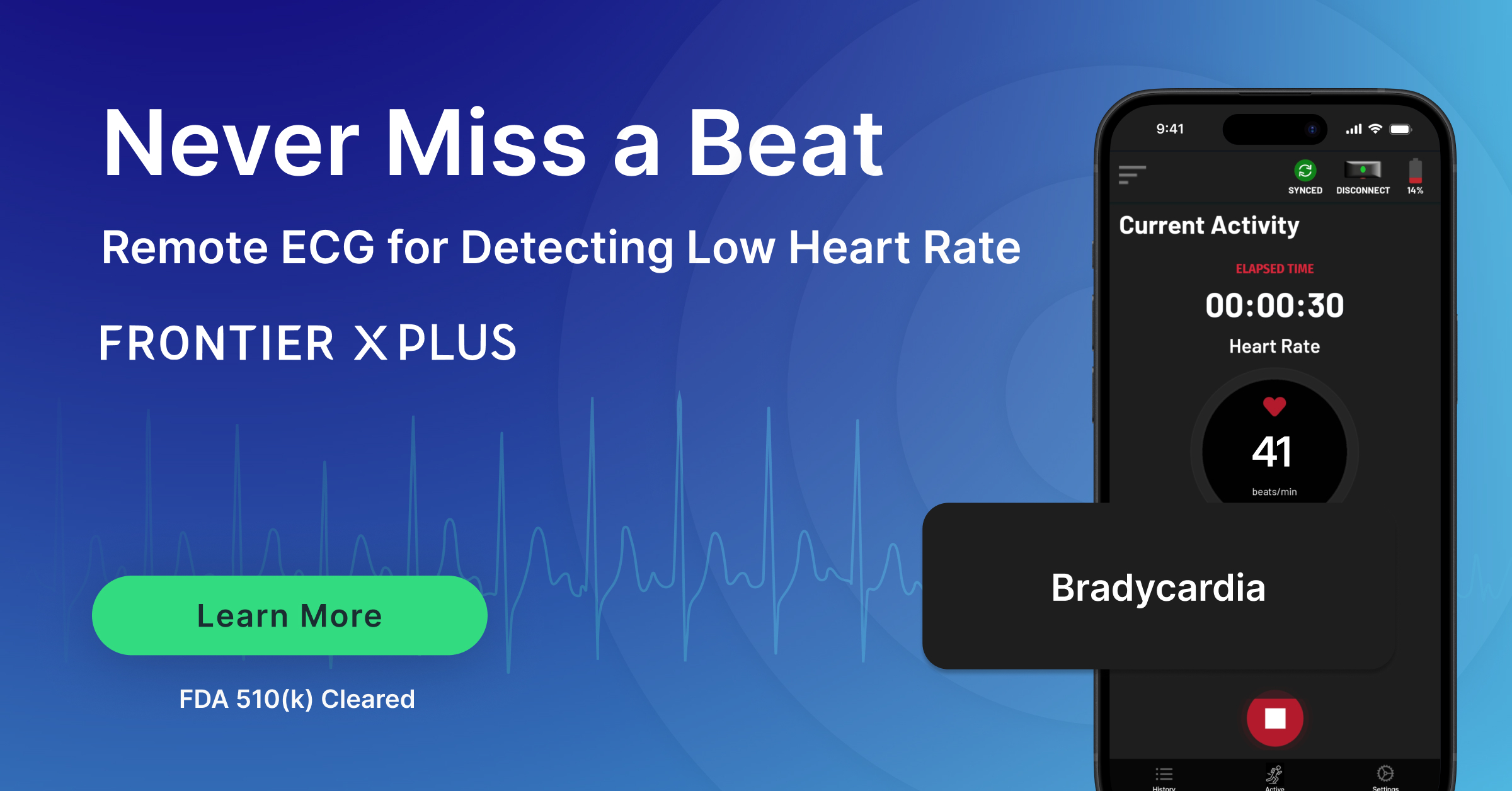
Your heart is more than a symbol of emotion – it’s a finely tuned muscle responsible for pumping blood throughout your body with precision. A normal resting heart rate for adults typically falls between 60 to 100 beats per minute (bpm). But what happens when your heart beats too fast or too slow? That’s where conditions like tachycardia and bradycardia come in.
In this blog, you’ll explore what these terms mean, what your heart rate might be telling you, and how ECG monitoring tools can help detect silent or intermittent heart rhythm issues before they become serious.
When Slower Isn’t Always Better
Take Olympic swimmer Mark Spitz, for example. Known for his elite athleticism, Spitz was diagnosed with bradycardia in 2018. Despite being in peak physical shape, he experienced symptoms after intense training sessions. What seemed like fatigue turned out to be a dangerously low heart rate. With early detection and proper monitoring, he was able to adjust his routine and manage his condition, proving that even champions aren’t immune to heart arrhythmias.
Spitz’s experience is a powerful reminder that a slow heart rate isn’t always a sign of fitness – sometimes, it’s a sign to pay attention. With the right tools and awareness, even hidden rhythm issues can be uncovered and managed before they become serious.
What Is Bradycardia?
Bradycardia is the opposite – a slower-than-normal resting heart rate, typically under 60 bpm. For some people, especially athletes, a slow heart rate can be a sign of strong cardiovascular health. But if your heart is beating too slowly to supply enough oxygen-rich blood to your body, it may result in:
- Fatigue
- Dizziness or lightheadedness
- Shortness of breath
- Fainting (syncope)
Bradycardia can be caused by issues in the heart’s natural pacemaker (the SA node), medication side effects, or underlying heart disease. Just like with tachycardia, not all cases are symptomatic, making it essential to track heart rhythms over time.
What Is Tachycardia?
Tachycardia refers to a resting heart rate that’s higher than normal, generally over 100 bpm in adults. It’s like your heart is revving its engine even when you’re parked. While it can be a normal response to exercise, stress, or excitement, persistent or unexplained tachycardia can signal an underlying issue.
There are several types of tachycardia:
- Ventricular tachycardia (VT): Starts in the lower chambers and can be life-threatening if not treated.
- Supraventricular tachycardia (SVT): A fast heart rate that begins above the ventricles, in the atria.
Some people feel symptoms like palpitations, chest discomfort, or shortness of breath. Others may not notice anything at all, which is why monitoring is so important.
Quick Guide: Heart Rate Ranges & What They Might Mean
| Heart Rate (bpm) | Classification | Possible Implications |
| Below 60 | Bradycardia | May be normal in athletes; could signal SA node dysfunction if symptomatic |
| 60–100 | Normal | Healthy resting heart rate for most adults |
| 100–130 | Mild Tachycardia | Could result from stress, fever, dehydration, physical activity or stimulant use |
| 130–160+ | Severe Tachycardia | Could result from intense exercise. May also indicate arrhythmia like SVT or VT- medical evaluation recommended |
Note: Heart rate should always be interpreted in the context of age, activity level, and symptoms.
Why Does Heart Rate Matter?
Your heart rate is a window into your cardiovascular health. While momentary fluctuations are normal, consistently high or low rates can lead to complications if left unchecked, including:
- Blood clots
- Heart failure
- Stroke
- Sudden cardiac arrest
What makes both tachycardia and bradycardia tricky is their potential to appear intermittently, especially during sleep, rest, or physical activity. These episodes may be missed by standard in-clinic ECGs or short-term Holter monitors.
Tachycardia and Bradycardia in Context: Beyond Resting Heart Rate
It’s important to note that both tachycardia and bradycardia aren’t limited to resting heart rate patterns alone. These rhythms can fluctuate during sleep, stress, hydration changes, or while transitioning between rest and exertion. For instance, inappropriate sinus tachycardia – where the heart races without clear triggers – and paroxysmal bradycardia – brief drops in heart rate that may cause fainting – are both challenging to catch with standard checkups. ECG monitoring bridges this gap by capturing these rhythm changes dynamically, offering clinicians more complete diagnostic insight into when and why these fluctuations occur.
When the Heart Swings: Alternating Bradycardia and Tachycardia
Some individuals may experience a condition known as tachy-brady syndrome, where the heart alternates unpredictably between too-slow and too-fast rhythms. This is particularly common in patients with sick sinus syndrome, a dysfunction of the heart’s natural pacemaker. These erratic swings can go unnoticed for years, especially if symptoms are mild or sporadic – such as occasional fatigue, brain fog, or palpitations. However, the consequences can be serious, including syncope or even sudden cardiac arrest. ECG monitoring is vital in capturing these transitions and enabling timely interventions, such as medication adjustments or pacemaker implantation if needed.
How ECG Monitoring Helps
This is where patchless ECG monitors come into play. Worn comfortably on the chest, it records high-quality ECG data in near-real-time, whether you’re working, resting, or exercising.
With the ECG monitors, you can:
- Capture both sinus rhythm and abnormal heart patterns like arrhythmia, SVT, or ventricular tachycardia.
- Tag symptoms during episodes for better physician interpretation.
- Sync data via a companion app for remote clinician access.
- Receive end-of-study reports reviewed by cardiac physiologists or your care team.
Unlike smartwatches that rely on 30–60 second spot checks, ECG monitors continuously record data for up to 24 hours per charge – making it much more effective at identifying silent or intermittent arrhythmias.
Who Should Be Monitoring Their Heart Rate?
You don’t have to wait for symptoms to take control of your heart health. ECG monitoring is especially helpful for:
- Individuals with known or suspected atrial fibrillation (AF)
- Those experiencing fluttering in the heart or palpitations
- Patients recovering from cardiac procedures
- Athletes pushing through endurance training
- Anyone with a family history of cardiovascular arrhythmia or abnormal heart rhythm
When to Talk to Your Doctor
If you’ve experienced:
- Skipped or irregular beats
- Rapid heart rate without exertion
- Lightheadedness or fatigue
- Nighttime chest discomfort or palpitations
It’s time to speak with your doctor. They may recommend a clinical-grade EKG/ECG machine or wearable electrocardiogram monitors for home use.
Final Thoughts
Your heart rate isn’t just a number – it’s a message from your body. Conditions like tachycardia and bradycardia might seem mild or go unnoticed at first, but over time, they can lead to serious outcomes. The good news? With advancements in ECG monitor technology, you can catch these issues early and take action.
Whether you’re trying to understand an odd flutter in your chest, monitor your atrial fibrillation heart rate, or manage a known condition, tools like ECG monitors provide clarity and control. They bridge the gap between how you feel and what your heart is actually doing – 24/7.
Don’t wait for a scary symptom to listen to your heart. Start monitoring today, and give your heart the attention it deserves.
Even the Fittest Can Miss the Signs
Atrial fibrillation doesn’t discriminate – it affects everyone from retired teachers to celebrities and world-class athletes. Basketball legend Larry Bird continued competing through dizzy spells later identified as AFib. Olympic swimmer Mark Spitz, despite his elite fitness, was blindsided by the diagnosis after a post-workout episode. Even public figures like former US presidents Joe Biden and George HW Bush, Arnold Schwarzenegger, and Susan Lucci have spoken openly about living with atrial fibrillation. What connects them all isn’t just their status — it’s the fact that AF often creeps in silently, revealing itself only through persistent monitoring. Their stories underscore an important truth: staying fit doesn’t mean you’re immune to cardiac arrhythmias, and that’s exactly why continuous ECG/EKG monitors are important for uncovering what you can’t always feel.
These stories underscore a powerful truth: staying fit doesn’t mean you’re immune to cardiac arrhythmias. Continuous ECG/EKG monitors are critical for uncovering what you can’t always feel.
If you’re someone who pushes hard during a run, a bike ride, or even a dance class — and feel the occasional flutter, breathlessness, or moment of fatigue — don’t ignore it. You might feel strong, but your heart could be telling a different story. That’s where the continuous EKG monitoring technology gives you power: real insight when you need it most.
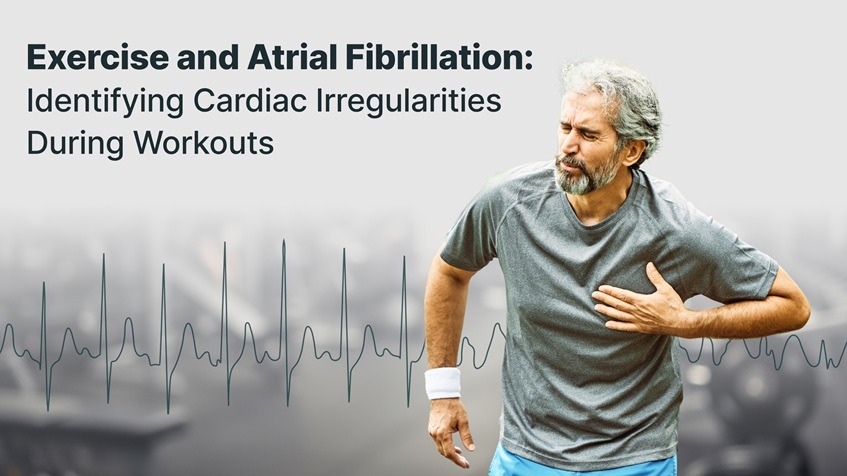
Why Exercise Can Trigger or Reveal Arrhythmias
Exercise can trigger arrhythmias by placing acute stress on the heart’s electrical system through increased sympathetic activity, elevated heart rate, and electrolyte imbalances from sweating. In individuals with genetic predispositions or structural heart conditions, this stress may provoke arrhythmias such as atrial flutter, supraventricular tachycardia (SVT), or even ventricular tachycardia
Additionally, sudden vagal rebound after intense workouts may destabilize the heart rhythm, particularly in endurance athletes. While exercise is beneficial for most, these physiological changes can unmask hidden electrical instability, making continuous ECG monitoring during and after exercise a critical tool for early detection.
Research indicates that overtraining or prolonged high-intensity activity may lead to atrial remodeling and increase the risk of AF over time — especially in athletes who have participated in endurance sports for many years. Thus, identifying early signs through wearable tech can prevent more serious episodes.
Symptoms to Watch During or After Exercise
If you have an underlying arrhythmia, symptoms may appear only when your heart is under pressure. These include:
- Shortness of breath
- Unexplained fatigue
- Rapid, irregular, or pounding heartbeat
- Dizziness or lightheadedness
- Chest discomfort or palpitations
Some people feel just a flutter; others may feel nothing at all. These subtle signs could point to atrial fibrillation or other heart irregularities that require evaluation
Unfortunately, Around one-third of those with AFib may not experience symptoms. — highlighting the importance of vigilance.
Who’s at Risk for Exercise-Triggered Arrhythmias?
- Aged adults — especially those with hypertension or prior history of cardiovascular arrhythmia
- Endurance athletes with long training histories (marathoners, triathletes, cyclists)
- Individuals with diagnosed Atrial Fibrillation (AF) with rapid ventricular response
- People with a family history of AFib or inherited arrhythmia syndromes
- Those experiencing increased palpitations, chest pressure, or breathing difficulty post-exercise
Long-term endurance sport practice is linked to a higher incidence of lone AF in men.
The Role of Continuous ECG Monitoring in Fitness
Traditional 12-lead ECG tests or short-term Holter monitors rarely capture arrhythmias that occur during physical exertion or recovery. That’s where continuous ECG monitoring becomes essential for active individuals.
These monitors offer:
- Continuous detection of AF events, bradycardia, and tachycardia
- Symptom tagging during workouts for contextual insight
- Instant syncing of ECG data to the app upon device connection, enabling timely clinician review.
Unlike smartwatches that offer brief 30-second spot checks, ECG monitors like the FDA-cleared, prescription-based Frontier X Plus and wellness-based Frontier X2 provide continuous, high-fidelity ECG data. This allows physicians to overread the recordings and detect arrhythmias that may occur during physical exertion – events often missed by short-duration monitors..
Key features include:
- Patchless and sweat-resistant, making it ideal for prolonged use during physical activity
- Delivers high-fidelity ECG data continuously, even during intense workouts
- Provides clinically reviewed summaries post-monitoring (especially if you have been prescribed a Holter monitor)
- Beneficial for individuals with existing heart conditions, athletes in post-treatment recovery, or anyone experiencing unexplained symptoms during exertion
Continuous ECG monitoring helps:
- Enable early detect for silent AF episodes
- Empower users to manage their heart health proactively
Empowering the Fitness-Minded With Better Tools
Whether you’re training for a race or simply trying to stay fit, ECG monitoring should not be an afterthought. Continuous ECG/EKG devices provide an unprecedented view into your cardiovascular rhythms — delivering actionable insights that you and your doctor can use to guide your care.
Athletes who report fluttering, skipped beats, or fatigue but receive “normal” clinic ECGs may simply not be monitored at the right time — typically when they’re resting. This is precisely why capturing data during and immediately after exertion is essential.
Moreover, for patients recovering from procedures like ablation or managing chronic AF through medication, these devices serve as a personalized health companion. They make it easier to recognize if interventions are working or if further changes are needed.
Final Thoughts
Exercise-induced arrhythmias are more common than you may think — and they often go unnoticed without the right monitoring. Whether you’re managing AF, atrial flutter, VT, SVT, or any form of EKG-detected arrhythmia, being proactive is key.
Wearable ECG monitors help bridge the gap between exercise and detection, allowing active individuals to train smarter, protect their long-term cardiovascular health, and gain peace of mind.
If you’ve experienced any irregular symptoms during workouts, or want to monitor your heart rhythm more closely, speak to your physician about using a continuous ECG/EKG device.
When it comes to heart health, there’s one truth many people don’t realize until they experience it firsthand: the rhythm of your life often mirrors the rhythm of your heart.
If you’ve been diagnosed with atrial fibrillation (AFib) – the most common atrial arrhythmia – you know how unsettling it can be. AFib causes the upper chambers of your heart (the atria) to quiver chaotically instead of beating in a steady, sinus rhythm. This leads to an irregular and often rapid heart rate, creating sensations like palpitations, a pounding heartbeat, dizziness, or even chest discomfort.
While medications and procedures like ablation play a critical role in treatment, many people overlook a key part of managing AFib: understanding and controlling personal lifestyle triggers.
In this blog, we’ll break down how daily habits – especially alcohol, caffeine, stress, and sleep patterns – may be silently influencing your heart rhythm. We’ll also explain how long-term ECG monitoring can help you identify your unique AFib patterns and make more informed choices to protect your heart health.
Why Triggers Matter in AFib Management
AFib isn’t always constant. For many people, it’s paroxysmal, meaning episodes come and go. The tricky part is that the triggers aren’t the same for everyone. Some people might notice their AFib symptoms right after a glass of wine, while others experience irregular beats following a stressful day or poor sleep.
The more you understand what sets off your episodes, the more control you can regain over your daily life. By identifying triggers early, you can:
- Reduce AFib burden (how often you experience episodes)
- Avoid progression to persistent or paroxysmal AFib
- Lower the risk of stroke, heart failure, and other complications
- Improve quality of life by reducing palpitations and discomfort
Alcohol and AFib: What the Research Shows
Does drinking alcohol increase heart rate?
In short: yes – for many people, it does.
Alcohol is one of the most well-documented AFib triggers. Studies show that even small amounts of alcohol can raise the risk of an AFib episode. In fact, the term “Holiday Heart Syndrome” was coined decades ago after doctors noticed a spike in arrhythmias during holiday seasons when people tend to drink more.
How Alcohol Affects Heart Rhythm
- Alcohol alters electrical signals in the atria, making it easier for chaotic rhythms to develop.
- It can cause dehydration, which throws off your body’s electrolyte balance – critical for a normal heartbeat.
- Alcohol may increase blood pressure, compounding the strain on your heart.
- Over time, heavy drinking can lead to cardiomyopathy, weakening the heart muscle itself.
What About Moderate Drinking?
Even moderate consumption – such as one drink a day – can raise your AFib risk. A meta-analysis published in Frontier of Cardiovascular Medicine found that a 1 drink/day increase in alcohol consumption increased the risk of AF by 6%
If you have AFib, doctors generally recommend limiting or avoiding alcohol. But the best way to know how it affects you personally is by tracking your episodes in real time.
Caffeine and AFib: Friend or Foe?
Coffee and high blood pressure have long been associated, but what about caffeine and AFib?
This is where things get a little more complicated. For years, doctors advised patients with atrial arrhythmia to avoid caffeine entirely. However, recent research suggests the link may not be as straightforward.
What the Studies Say
- Some people are sensitive to caffeine, and even a small amount can cause palpitations or an erratic heart rhythm.
- For others, moderate coffee intake may actually be neutral or even protective for the heart.
- Energy drinks, however, often contain very high levels of caffeine combined with other stimulants and have been linked to dangerous ventricular activity and arrhythmias.
Bottom Line on Caffeine
If you’ve noticed that your heart feels like it is racing after coffee or tea, it’s worth experimenting by cutting back. Keep in mind that continuous heartmonitoring can help determine whether your ventricular rhythm types or atrial rhythm patterns are actually changing after caffeine intake.
Stress and Sleep: The Silent Contributors
Stress and atrial fibrillation are closely linked. Emotional stress triggers a surge of catecholamines – hormones that speed up your heart rate and can tip your system into AFib.
Poor sleep is another hidden culprit. Conditions like sleep apnea or insomnia increase your risk of AFib by contributing to heart strain, spikes in blood pressure, and changes in heart rhythm during the night.
How to Track the Impact
- Keep a symptom journal noting stressful events, restless nights, or panic episodes.
- Use wearable ECG monitors that track your heart rate during sleep and heart rhythm at rest to identify nighttime disturbances.
How Long-TermECG Monitoring Helps You Stay Ahead
Standard checkups or Holter exams only offer a snapshot of your heart’s activity. But atrial fibrillation could often happen outside the doctor’s office.
That’s why long-termECG monitoring devices are becoming an essential part of AFib management. These wearable devices:
- Record both sinus rhythm and arrhythmias over 24 hours or longer
- Help correlate symptoms like palpitations with real ECG data
- Allow you to see how specific activities (like drinking alcohol or exercising) affect your heart rhythm
- Provide detailed reports for your healthcare provider to adjust treatment
Unlike basic fitness trackers, ECG monitors that record continuously, such as the Frontier X Plus, provide high-fidelity ECG and detect Afib episodes.
Practical Tips to Reduce Your AFib Triggers
Managing AFib isn’t just about medications or heart failure treatment – it’s also about lifestyle. Here’s how you can reduce your episodes:
- Limit or avoid alcohol, especially binge drinking.
- Monitor your caffeine intake. May try decaf or reduce portion sizes if you’re sensitive.
- Prioritize good sleep hygiene. Treat sleep apnea if diagnosed.
- Manage stress with relaxation techniques like yoga, meditation, or breathing exercises.
- Track your episodes with a heart rhythm monitor to spot personal patterns.
- Discuss findings with your doctor to adjust your AFib treatment plan.
Final Thoughts
Understanding your personal AFib triggers – whether it’s drinking and atrial fibrillation, caffeine and AFib, or emotional stress – empowers you to live a fuller, healthier life. By pairing lifestyle awareness with tools like long-termECG monitoring, you can make informed decisions that help protect your heart health and reduce the burden of atrial fibrillation.
Whether you’re dealing with occasional palpitations or frequent arrhythmia episodes, tracking your heart rhythm daily gives you and your doctor the data needed to adjust care and reduce future risks.
FAQs
Can alcohol cause AFib to start suddenly?
Yes. For many people, even a single drink can trigger a sudden episode of atrial fibrillation, especially if they are already predisposed.
Does coffee increase AFib risk?
Caffeine sensitivity varies. Some people find that coffee causes palpitations, while others do not experience any effect on their heart rhythm.
How can I monitor my AFib triggers at home?
Using a long-termECG monitor allows you to track your heart rhythm throughout the day and correlate arrhythmia episodes with specific triggers like alcohol, caffeine, or stress.
What is the best treatment for AFib triggered by lifestyle factors?
In addition to AFib medications, lifestyle modifications such as reducing alcohol, managing stress, and improving sleep can significantly reduce episodes.
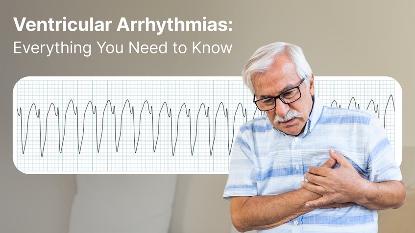
When your heart beats too fast, too slow, or erratically, it can feel like something is off. If you’ve ever thought, “My heart feels like it is racing,” or experienced a sudden, rapid heartbeat out of nowhere, you’re not alone. For many people, these sensations may be signs of ventricular arrhythmias, a group of heart rhythm disturbances originating in the lower chambers of the heart – the ventricles.
These conditions range from premature beats to potentially life-threatening rhythms like ventricular tachycardia (V Tach) and ventricular fibrillation (V Fib). Understanding these irregular rhythms is critical because early detection and management can prevent severe complications, including sudden cardiac arrest.
This comprehensive guide breaks down everything you need to know about ventricular arrhythmias, from their causes to treatment options and the latest in monitoring technology.
What Are Ventricular Arrhythmias?
Ventricular arrhythmias refer to abnormal electrical signals in the heart’s ventricles – the two lower chambers responsible for pumping blood to the lungs and the rest of the body. These disturbances can lead to inefficient blood flow, resulting in symptoms like dizziness, palpitations, fainting, or even cardiac arrest.
Types of Ventricular Arrhythmias
Understanding ventricular rhythm types is essential because not all ventricular arrhythmias are equally dangerous. Here’s a breakdown:
- Premature Ventricular Contractions (PVCs): They are early, extra heartbeats that start in the heart’s lower chambers (the ventricles), disrupting the heart’s normal rhythm for a moment
- Ventricular Tachycardia (V Tach): It is a rapid heart rhythm – more than 100 beats per minute – that begins in the lower chambers of the heart. This condition prevents the heart’s chambers from filling adequately with blood, reducing the amount that is pumped to the body.
- Ventricular Fibrillation (V Fib): is a life-threatening heart rhythm where the ventricles quiver or fibrillate chaotically instead of expanding and contracting effectively. This prevents the heart from pumping out blood. Within seconds, blood flow to the brain stops, causing loss of consciousness. Without immediate treatment, usually with defibrillation, V-Fib leads to sudden cardiac arrest.
Symptoms of Ventricular Arrhythmias
Many patients describe ventricular arrhythmias as feeling like a “sudden rapid heartbeat” or an erratic heart rhythm. Other common symptoms include:
- Chest pain or discomfort
- Dizziness or fainting (syncope)
- Shortness of breath
- Palpitations or fluttering sensations in the chest
- Fatigue or weakness
These symptoms often trigger a search for a heart monitor to check for arrhythmia, which helps confirm the diagnosis.
What Causes Ventricular Arrhythmias?
Several factors can lead to abnormal ventricular activity:
- Coronary artery disease
- Cardiomyopathy (weak or enlarged heart muscle)
- Heart failure
- Previous heart attack (myocardial infarction)
- Electrolyte imbalances
- Genetic conditions like arrhythmogenic right ventricular cardiomyopathy (ARVC)
- Medications or stimulant abuse
- Structural heart changes
In some cases, the cause remains unknown – a condition called idiopathic ventricular arrhythmia.
Why Are Ventricular Arrhythmias Dangerous?
Ventricular arrhythmias are a leading reason for cardiac arrest, especially when they progress to V Fib. The rapid and disorganized contractions prevent the heart from pumping blood, cutting off oxygen to the brain (cardiac arrest) and other vital organs. Without immediate intervention, this can lead to death within minutes.
Even if life-threatening events are avoided, heart rhythm problems like sustained V Tach can weaken the heart over time, increasing the risk of heart failure and other complications.
Diagnosing Ventricular Arrhythmias
If you suspect a ventricular rhythm issue, healthcare providers will often recommend a combination of tests:
Common Diagnostic Tools:
- Electrocardiogram (ECG):
The first-line test to detect arrhythmias in real-time. - Medical Holter Monitor:
A portable device worn for 24 to 48 hours to catch intermittent heart rhythm disturbances. - Event Monitors & Loop Recorders:
Long-term devices for patients with infrequent but concerning symptoms. - Echocardiogram & Cardiac MRI:
Used to check for structural abnormalities contributing to arrhythmias. - Electrophysiology Study (EPS):
A specialized test to map the heart’s electrical pathways and identify the source of abnormal signals. - Exercise stress test : if symptoms occur under exertion, exercise stress tests will often be ordered to look for these conditions
Treatment Options for Ventricular Arrhythmias
Treatment depends on the type and severity of the arrhythmia, symptoms, and underlying causes.
Medications
- Antiarrhythmic Drugs:
These are prescribed to stabilize ventricular activity. Common ventricular tachycardia treatment drugs include amiodarone and lidocaine. - Beta-Blockers:
Reduce the risk of dangerous arrhythmias, especially in patients with heart disease. - Calcium Channel Blockers:
Sometimes used in specific arrhythmia cases.
Catheter Ablation
For frequent or life-threatening V Tach, doctors may recommend ventricular tachycardia ablation. This minimally invasive procedure targets and destroys the tissue responsible for the abnormal electrical signals.
Ventricular Tachycardia Ablation Success Rate:
Recent studies show that catheter ablation has a success rate of around 70-80% in controlling V Tach, especially in patients with structurally abnormal hearts.
Implantable Cardioverter-Defibrillator (ICD)
An ICD is a device implanted under the skin that detects abnormal ventricular rhythms and delivers shocks to restore normal rhythm. ICDs are life-saving for people at high risk of sudden cardiac death.
The Role of Continuous Monitoring
Continuous monitoring plays a pivotal role in diagnosing and managing ventricular arrhythmias. Unlike standard ECGs that capture a snapshot in time, ECG monitors that record continuously over many hours or days, provide high-fidelity ECG to doctors, thereby enabling them to detect:
- PVC patterns
- V Tach episodes
- Irregular heartbeats that are symptomless
These devices also empower patients by providing near-real-time insights into their heart health, ensuring that erratic heart rhythms don’t go unnoticed.
Prevention and Risk Reduction
While some ventricular arrhythmias are unavoidable due to genetics or prior heart damage, you can take steps to reduce your risk:
- Maintain a heart-healthy lifestyle
- Treat high blood pressure and cholesterol
- Avoid stimulant drugs and excessive caffeine
- Manage sleep apnea
- Follow your doctor’s advice if you have known heart diseases
- Follow an appropriate exercise plan
When to See a Specialist
If you’ve experienced symptoms like palpitations, dizziness, or episodes where my heart feels like it is racing, it’s crucial to consult a healthcare provider. A ventricular tachycardia specialist or electrophysiologist can help determine the cause and recommend the best treatment path.
Final Thoughts
Ventricular arrhythmias are more than just uncomfortable sensations – they can be serious indicators of underlying heart rhythm problems. From premature ventricular complexes to life-threatening V Fib, these conditions require prompt attention, proper diagnosis, and often long-term management.
With advancements in irregular heartbeat monitor technology, patients and physicians now have better tools to detect, monitor, and treat these rhythms early – before they lead to serious complications.
FAQs: Ventricular Arrhythmias
Q: What’s the difference between V Tach and V Fib?
A: In V Tach, ventricles contract in a coordinated manner but do so very rapidly. In V Fib, ventricles contract in an uncoordinated manner.
Q: What is the success rate of V Tach ablation?
A: Ventricular tachycardia ablation has a success rate of 70-80% for reducing arrhythmia episodes.
Q: How do I know if my rapid heartbeat is dangerous?
A: Persistent sudden rapid heartbeat with symptoms like fainting or chest pain requires immediate medical attention.
If you’re concerned about ventricular arrhythmias, talk to your healthcare provider about continuous monitoring options to keep your heart health a priority.
The cruel trick of arrhythmias: silence while you rest, symptoms when you rise.
When it comes to heart health, arrhythmias don’t always follow the rules. At rest, you could be reading a book, working at your desk, or even asleep – yet your heart may be slipping into an irregular rhythm without you realizing it. Later, when you exercise, climb stairs, or push yourself a little harder, that same hidden rhythm can suddenly make itself felt – palpitations, dizziness, or even fainting.
If you’ve ever wondered why your heart skips a beat, why you feel drained for no clear reason, or whether your racing heartbeat is something more than stress, you’re not alone. Many people face these questions without answers.
What an Arrhythmia Really Means for You
An arrhythmia is simply an irregular rhythm of your heartbeat. But “simple” doesn’t mean harmless. Some arrhythmias may cause nothing more than the occasional skipped beat. Others, like atrial fibrillation (AFib) or ventricular tachycardia, carry serious risks.
The most common arrhythmias include:
- Atrial fibrillation (AFib): The most common sustained arrhythmia. You may not feel it, yet it raises your risk of stroke fivefold.
- Supraventricular tachycardia (SVT): A sudden racing heartbeat that can feel alarming, even if brief.
- Ventricular tachycardia (VT): A rhythm disturbance in the heart’s lower chambers that can be life-threatening.
- Bradycardia: When your heartbeat slows too much, leaving you dizzy, faint, or constantly fatigued.
The Silent Arrhythmias You Might Never Notice
The crucial part of some arrhythmias is their occurrence without any symptoms. You may feel perfectly fine, while your heart is misfiring in the background. You might go years without a single symptom, but your risk of stroke or other complications grows quietly in the meantime.
This is especially true with atrial fibrillation. Many people discover it only after a major event. Imagine your heart “screaming” on the inside, but your body giving you no clue. That’s what makes silent arrhythmias so dangerous.
When Symptoms Hit You During Activity
On the flip side, you might feel absolutely normal at rest – only to have symptoms appear during activity. Maybe it’s while jogging, playing a game, or even walking quickly to catch a train. Suddenly, your heart races, your chest feels tight, or you’re left gasping for air. No wonder cases like Juan Izquierdo, the 27-year-old footballer who collapsed during a Copa Libertadores match, resonate so strongly. Despite being diagnosed years earlier with a ‘small arrhythmia,’ he had no symptoms—until exertion pushed his heart into crisis.
These episodes aren’t just unpleasant – they can be dangerous. Exercise-induced arrhythmias, such as ventricular tachycardia, can escalate quickly. If you’ve ever brushed off palpitations or dizziness as “just pushing too hard,” it’s worth taking a closer look.
Why the Typical Tests Often Fail You
Here’s the frustrating part: most medical tests look at your heart for only a short time. A 10-second ECG in your doctor’s office, or even a 24-hour Holter monitor, might completely miss your arrhythmia.
Why? Because your heart may not always misfire when it’s being watched. The skipped beats may happen while you’re asleep, or during a random workout two days later. Short-term checks give you snapshots, but what you need is the full story.
This leaves you with questions:
- Was that flutter I felt just stress – or something serious?
- Am I safe to keep exercising?
- Could I be living with a silent arrhythmia and not know it?
How Continuous Monitoring Changes the Game
The only way to truly understand your heart is to see what it’s doing across your whole life – not just for a few minutes in a clinic. That’s where ECG monitors that record continuously come in.
With devices like:
- Frontier X Plus (a prescription-based, FDA-cleared device for medical use)
- Frontier X2 (a wellness-focused monitor for athletes and health-conscious individuals)
– you can track your ECG continuously. Whether you’re sitting at your desk, walking your dog, or pushing through a workout, every beat is captured.
For you, this means:
- No more guessing: You’ll know if your dizziness or fatigue is linked to your heart.
- Peace of mind: Silent arrhythmias won’t go unnoticed.
- Better decisions: If you’re an athlete, you can train with confidence. If you’re managing a condition, your doctor can access your data via a cloud and an app and guide treatment.
The Real Risks You Face
Missing arrhythmias comes with a huge cost:
- Silent AFib increases your risk of stroke, even if you never feel a symptom.
- Exercise-induced arrhythmias can trigger sudden cardiac events.
- Quality of life suffers when unexplained fatigue or palpitations leave you anxious.
- Athletes risk pushing their hearts past safe limits without knowing it.
It’s not about fear – it’s about being informed. Your heart shouldn’t be a mystery.
Why You Need to Pay Attention Now
If you’ve ever brushed off chest flutter, ignored fatigue, or chalked up dizziness to dehydration, it may be time to take your heart health seriously. You don’t want to wait until a missed arrhythmia leads to something worse.
The paradox is clear: your heart could be quiet when you expect noise, or noisy when you expect quiet. The only way to know for sure is to listen more closely – every beat, every day.
Join Us Live: Learn From the Experts
This AFib Awareness Month, we’re bringing you insights from two leading Cleveland Clinic cardiologists:
Silent to Symptomatic: Arrhythmias Across Daily Life and Exercise
Thursday, September 25, 2025
9:00–10:00 AM EDT
6:30–7:30 PM IST
Speakers
- Dr. Niraj Varma – Professor of Medicine, Section of Electrophysiology and Pacing, Cleveland Clinic
- Dr. Tamanna Singh – Co-Director, Sports Cardiology Center, Cleveland Clinic
They’ll break down how arrhythmias appear in rest vs exercise, what you need to watch for, and how continuous monitoring can protect your heart.
FAQs on Arrhythmias and Heart Health
Q1. Could I really have an arrhythmia without knowing it?
Yes. Many arrhythmias, especially atrial fibrillation, can be silent. You may feel perfectly fine while your heart is in an irregular rhythm.
Q2. What symptoms should I watch out for?
Pay attention to palpitations, chest flutter, dizziness, fainting, unexplained fatigue, or shortness of breath. These may be signs of an irregular heart arrhythmia.
Q3. What are the most common arrhythmias?
The most common include atrial fibrillation, supraventricular tachycardia, ventricular tachycardia, and bradycardia.
Q4. How are arrhythmias treated?
Treatment depends on the type. It may involve medication, ablation procedures, implantable devices, or lifestyle adjustments.
Q5. Why is atrial fibrillation so serious?
Because it raises your risk of stroke. Even if you never feel symptoms, AFib can cause clots that travel to the brain.
Q6. How does long-termECG monitoring help me?
It captures your heartbeat during your real life – while you rest, work, and exercise. This way, silent arrhythmias don’t go unnoticed, and symptomatic ones are tracked with context.
Q7. Should I consider long-term monitoring?
If you’ve had unexplained palpitations, fainting, or fatigue – or if you’re an athlete returning to training after a cardiac scare – yes, long-term monitoring may be right for you.
Final Takeaway
Your heart doesn’t always play by the rules. It can be silent when you expect noise, and noisy when you expect silence. That paradox makes arrhythmias easy to miss – but dangerous to ignore.
With long-termECG monitoring and expert care, you can finally uncover what your heart is really doing. Don’t wait for symptoms to decide your future – listen to your heart now.
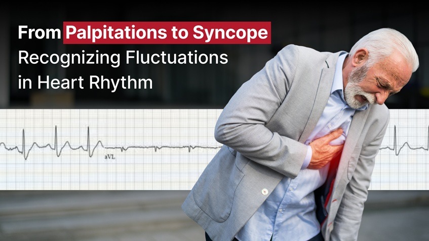
Our hearts beat thousands of times per day, a rhythmic cadence that most of us never notice. But for some, that cadence occasionally stumbles, quickens, or falters. These irregularities in heart rhythm can manifest as palpitations or even lead to sudden episodes of fainting, known as syncope. Understanding what these symptoms mean and when they warrant medical attention is crucial for protecting your cardiovascular health.
In this blog, we’ll explore the spectrum of heart rhythm abnormalities – from harmless skips to potentially serious arrhythmias – and explain how long-term ECG monitoring can play a vital role in early detection and prevention.
What Are Palpitations?
Palpitations are sensations of a rapid, fluttering, or pounding heart. Some people describe them as skipped beats or a flip-flop feeling in the chest. These sensations can occur during physical activity, emotional stress, or even while at rest.
Common Causes of Palpitations:
- Premature atrial or ventricular contractions (PACs or PVCs)
- Anxiety or panic attacks
- Caffeine or stimulant intake
- Dehydration or electrolyte imbalance
- Thyroid disorders
- Anemia or low blood pressure
In many cases, palpitations are benign and don’t indicate a serious problem. However, persistent or recurrent palpitations may signal a more serious underlying arrhythmia.
When Palpitations Are a Warning Sign
While occasional palpitations can be harmless, certain red flags increase the likelihood that they are associated with a cardiac rhythm disorder:
- Palpitations accompanied by dizziness or fainting
- Chest pain or shortness of breath
- Palpitations that last more than a few minutes
- A resting heart rate above 100 bpm without clear cause
If you experience any of the above, it’s essential to seek evaluation. These could indicate supraventricular tachycardia (SVT), atrial fibrillation (AFib), or ventricular tachycardia (VT) – conditions that may require treatment.
What Is Syncope?
Syncope is the medical term for a temporary loss of consciousness, commonly referred to as fainting. It occurs when there is a sudden drop in blood flow to the brain. While syncope can be caused by non-cardiac conditions (like a vasovagal response or dehydration), cardiac syncope is often the most concerning.
Cardiac Causes of Syncope:
- Arrhythmias (bradycardia, tachycardia, pauses)
- Structural heart disease (aortic stenosis, hypertrophic cardiomyopathy)
- Heart block (electrical conduction delays)
- Prolonged QT syndrome
Sudden syncope without warning – especially during exercise or while lying down – is more likely to be cardiac in nature and should never be ignored.
The Spectrum of Heart Rhythm Abnormalities
Understanding where your symptoms fall on the arrhythmia spectrum can help guide evaluation and treatment:
1. PACs and PVCs
These are early beats that originate in the atria or ventricles. They often feel like a skipped or extra beat and are usually benign unless frequent or occurring in a pattern.
2. Sinus Tachycardia
An elevated heart rate due to physiological triggers like exercise, anxiety, or fever. While not an arrhythmia per se, persistent sinus tachycardia may require investigation.
3. Atrial Fibrillation
A chaotic rhythm originating in the atria that leads to an irregular and often rapid heartbeat. AFib is a common cause of palpitations and increases stroke risk.
4. SVT (Supraventricular Tachycardia)
Sudden episodes of fast heart rate that often begin and end abruptly. SVT can cause palpitations, chest discomfort, and even syncope.
5. Ventricular Tachycardia (VT)
A potentially life-threatening rhythm originating in the ventricles. Symptoms include palpitations, dizziness, and syncope.
6. Bradycardia and Heart Block
Slow heart rhythms, often due to conduction system dysfunction. May lead to fatigue, dizziness, or syncope.
Why Timing and Pattern Matter
One of the challenges in diagnosing arrhythmias is their episodic nature. You may experience symptoms only a few times a week, or during sleep or exercise, making them hard to catch with a standard spot check ECG or even a short-term Holter monitor.
How Long-Term ECG Monitoring Helps
Long-term ECG monitors allow for long-duration recording of heart rhythm data. These devices are typically worn on the chest and are capable of capturing both normal and abnormal rhythms over days or weeks. Here’s how they help:
- Catch intermittent arrhythmias missed by standard ECGs
- Correlate symptoms (like palpitations or dizziness) with rhythm data
- Aid physicians in diagnosis and treatment planning
- Provide a timeline of events for better pattern recognition
ECG monitoring is especially valuable for individuals with unexplained syncope, palpitations, or those at high risk for arrhythmias due to family history or underlying cardiac conditions.
When Should You Be Concerned?
Here are some situations where ECG monitors could be life-saving:
- Frequent palpitations with no clear trigger
- Family history of sudden cardiac death
- Fainting during exercise or while lying down
- Heart rate that spikes or drops without exertion
- Known arrhythmia being treated with medications
Working with Your Doctor
Symptom diaries, paired with continuous ECG reports, give cardiologists or electrophysiologists a more complete picture. These tools allow them to:
- Diagnose arrhythmias more accurately
- Assess treatment efficacy
- Decide if interventions like ablation or pacemaker implantation are necessary
Conclusion: Don’t Dismiss the Flutter
Palpitations and syncope can range from harmless to serious. What makes them dangerous is unpredictability and the difficulty of diagnosing the underlying cause based on a short clinical snapshot. That’s why long-term ECG monitoring is a game changer.
If you’ve ever wondered whether that skipped beat was something more, or if you’ve felt dizzy or blacked out without explanation, don’t wait. Consult a medical professional, discuss monitoring options, and take charge of your heart health today.
Because when it comes to your heart, every beat matters.
Your heart is the engine of your body, tirelessly pumping blood and sustaining life. But when its rhythm is disrupted, especially in the form of tachycardia – an abnormally fast heartbeat – it can become more than just a flutter. Two major types of tachycardia include Supraventricular Tachycardia (SVT) and Ventricular Tachycardia (VT), each with distinct causes, implications, and treatments.
In this blog, we explore the difference between SVT and VT, their symptoms, risks, and how continuous ECG technology and modern treatment options are revolutionizing diagnosis and care.
What is Supraventricular Tachycardia (SVT)?
SVT refers to any tachyarrhythmia originating above the ventricles, typically in the atria (the upper chambers), but not originating in the sinoatrial node. These arrhythmias result in a heart rate that can soar to 150-220 beats per minute. SVT rhythm is usually regular; exceptions include atrial fibrillation (AFib) and multifocal atrial tachycardia (MAT).
Types of SVT:
- Atrial Flutter
- Atrial Fibrillation
- Premature atrial contractions (PACs)
- Focal atrial tachycardia
- Multifocal atrial tachycardia (MAT)
- Atrioventricular nodal reentrant tachycardia (AVNRT)
- Atrioventricular reciprocating tachycardia (AVRT)
Symptoms of SVT can be:
- Palpitations
- Lightheadedness
- Shortness of breath
- Chest discomfort
- Fatigue or anxiety
Triggers can be:
- Excess caffeine or alcohol
- Stress
- Sleep deprivation
- Certain medications
Read More: Understanding Supraventricular Tachycardia (SVT): Symptoms, Causes, and Treatment
What is Ventricular Tachycardia (VT)?
VT is a rapid heart rhythm that originates in the ventricles. Unlike SVT, VT can be considered as life-threatening because it affects the heart’s main pumping chambers. Left ventricular tachycardia, in particular, can severely reduce cardiac output, leading to dizziness, fainting, and even sudden cardiac arrest.
Types of VT:
- Sustained VT
- Non-sustained VT
- Monomorphic VT (consistent rhythm)
- Polymorphic VT (variable rhythm)
Common Symptoms can be:
- Dizziness
- Syncope (fainting)
- Weak pulse or no pulse
- Sudden cardiac arrest
Risk Factors can be:
- Previous heart attack
- Heart failure
- Dilated cardiomyopathy
- Congenital heart disease
VT causes most cases of sudden cardiac death, with an estimated rate of 300,000 deaths each year in the United States.
Read More: What is Ventricular Tachycardia and What Are The Most Common Signs?
Comparing SVT and VT: What Sets Them Apart?
| Feature | Supraventricular Tachycardia (SVT) | Ventricular Tachycardia (VT) |
| Origin | Above the ventricles | Within the ventricles |
| Heart Rate | 150-220 bpm | Often >100 bpm |
| Severity | rarely dangerous, unless they stay in SVT for too long | Potentially life-threatening |
| Common Triggers | Stress, caffeine, stimulants | Structural heart disease |
| Treatment Options | Vagal maneuvers, drugs for SVT, SVT ablation | VT drugs, defibrillation, VT ablation |
| Detection | ECG, Holter monitor | ECG, Holter monitor |
Why Does Differentiation Matter?
Correctly identifying whether a fast heart rhythm is SVT or VT can drastically change the course of treatment. Treating VT as SVT can delay life-saving interventions. On the other hand, overtreating SVT as a life-threatening emergency can cause unnecessary stress and medical procedures.
Both conditions require expert diagnosis, but the consequences of mismanagement differ greatly. Continuous heart monitoring is pivotal for capturing these arrhythmias, especially when they are intermittent or asymptomatic.
How Are These Conditions Diagnosed?
The first step is a 12-lead ECG, but due to the fleeting nature of SVT and VT episodes, continuous heart monitoring using medical Holter monitors or advanced wearable monitors provides invaluable insight.
Additional diagnostic tools include:
- Echocardiogram (to assess heart structure)
- Stress tests (to provoke arrhythmia under observation)
- Electrophysiology studies (to map abnormal electrical pathways)
Tests for SVT may involve:
- Event monitors
- Implantable loop recorders
- SVT monitors with patient-triggered symptom tagging
- If symptoms occur under exertion, exercise stress tests will often be ordered to look for these conditions
Treatment Pathways
SVT Treatment Options may be
- Vagal maneuvers as advised by doctors
- Medical treatment for SVT: Beta-blockers, calcium channel blockers
- Drugs for SVT as prescribed by your doctor
- SVT ablation: Catheter ablation procedures targeting the electrical misfiring
- Supraventricular tachycardia catheter ablation: High success rates and often considered first-line for recurrent episodes
Ventricular Tachycardia Treatment may be
- Drugs for VT as prescribed by your doctor
- Ventricular tachycardia treatment drugs: Antiarrhythmic agents tailored to the patient’s heart condition
- Implantable cardioverter-defibrillator (ICD): For high-risk patients
- Ventricular tachycardia ablation success rate: Can reach 70-90% depending on the heart’s structural integrity and underlying cause
- Emergency defibrillation: In cases of pulseless VT
Disclaimer: Medication should be based on a patient’s medical history, current medications, and other patient factors.
Role of Continuous Heart Monitoring
Early detection is essential. Symptoms can be misleading, and many arrhythmias are asymptomatic, especially in early stages.
With continuous heart monitoring:
- Episodes are captured in real-time, even while sleeping or exercising
- Heart rhythm monitors provide a full picture of both SVT and VT frequency
- Data-driven treatment becomes possible, improving outcomes

Prevention and Risk Reduction
While not all cases of SVT or VT are preventable, you can reduce your risk with healthy lifestyle choices and medical supervision:
Prevention Tips:
- Maintain a healthy weight and blood pressure
- Limit caffeine and alcohol
- Manage stress with mindfulness or therapy
- Avoid recreational drugs and smoking
- Get regular exercise, approved by your doctor
- If you have a family history, consider genetic testing or early screening
Final Thoughts
SVT and VT are both forms of tachycardia, but the stakes and strategies involved in treating them are different. The health of the heart depends not just on its strength but also on its rhythm.
Whether you’re managing an SVT heart condition or facing the risks of VT, timely diagnosis, continuous heart monitoring, and appropriate treatments are critical. From vagal maneuvers for SVT to ventricular tachycardia treatment drugs, modern medicine offers effective pathways to manage both.
ECG monitors that can record continuously have transformed the landscape for patients and clinicians alike – offering an accessible, non-invasive window into the heart’s electrical activity in real time.
FAQs
-
What is the main difference between SVT and VT?
SVT originates above the ventricles (usually in the atria), while VT originates in the ventricles. SVT is generally less dangerous than VT, which can lead to sudden cardiac arrest.
-
Can SVT turn into VT?
Not directly. However, individuals with predisposing conditions could experience both types of arrhythmias.
-
Is VT always life-threatening?
No, but sustained VT is a medical emergency and can lead to cardiac arrest if not treated immediately.
-
What are the treatment success rates for VT ablation?
Depending on the cause and patient profile, VT ablation success rates have been over 75%.
-
What is the best monitor to detect SVT or VT at home?
Continuous ECG monitors or medical Holter monitors are the most effective tools for detecting intermittent arrhythmias like SVT and VT.
-
Are there medications available for SVT and VT?
Yes. Drugs like adenosine and beta-blockers are used for SVT, while amiodarone and lidocaine are common for VT (Disclaimer: medication should be based on a patient’s medical history, current medications, and other patient factors)
-
What are vagal maneuvers?
Simple actions like coughing or bearing down can help interrupt SVT episodes and restore normal rhythm.
-
Can I live a normal life with SVT or VT?
With proper treatment and monitoring, many individuals with these arrhythmias can lead normal, active lives.
Have you ever checked your pulse and found it unusually slow, especially after a workout or first thing in the morning? For athletes and fitness enthusiasts, a resting heart rate(RHR) below 60 beats per minute (bpm) might be completely normal. But for others, it could be a warning sign. So how do you know whether a slow heart rate is healthy or something more serious?
In this blog, we’ll break down the differences between athletic bradycardia, sinus bradycardia, and general bradycardia, and explain how ECG monitoring with a tool like the FDA-cleared Frontier X Plus can help you stay confident and safe.
What Is Bradycardia?
Bradycardia is the clinical term for a heart rate below 60 bpm in adults. While that may sound concerning, it’s not always dangerous. Many people — especially trained athletes — naturally have a slower resting heart rate because their hearts are more efficient at pumping blood.
But not all bradycardia is created equal.
1. Athletic Bradycardia: A Sign of a Strong Heart
If you’re a runner, cyclist, swimmer, or just very active, you may notice that your RHR is below the range of 60 to 80 bpm for the average adult; a young, healthy athlete may have a heart rate of 30 to 40 bpm. This is called athletic bradycardia.
- Cause: Long-term endurance training boosts the strength of the heart muscle and increases vagal tone, which slows the heart rate.
- Rhythm: Normal sinus rhythm — just slower.
- Symptoms: None — athletes usually feel great.
This type of bradycardia is normal, harmless, and even desirable in the athletic population.
2. Sinus Bradycardia: When Slowness Starts at the Source
Sinus bradycardia refers to a slow heart rate caused by the sinoatrial (SA) node, your heart’s natural pacemaker. It may occur in people during sleep, at rest, or as a side effect of medications (like beta-blockers).
- Cause: Normal physiology, medications, or medical conditions
- Rhythm: Still a regular sinus pattern, but slow
- Symptoms: May be asymptomatic or cause fatigue, lightheadedness, or fainting
Sometimes normal, sometimes a signal for further evaluation — especially if symptoms are present.
3. Concerning Bradycardia: When Slow Is a Red Flag
Not all slow heart rhythms come from the SA node. Bradycardia may also result from electrical signal blockages in the heart, such as AV block or junctional rhythms. These cases often require intervention.
- Cause: Heart disease, aging, or damaged electrical pathways
- Rhythm: Irregular or abnormal patterns (not sinus)
- Symptoms: Fatigue, fainting (syncope), dizziness, confusion
These forms of bradycardia should be taken seriously and evaluated by a cardiologist.
Why It’s Hard to Tell the Difference
If you’re healthy and active, it’s easy to dismiss a slow heart rate as part of your fitness gains. But what if you’re not sure? Or what if symptoms show up during rest, sleep, or exercise? Traditional ECGs or smartwatches don’t give you the whole picture — especially if symptoms are intermittent.
That’s where ECG monitors become invaluable.
Read More : How Frontier X Plus is Better Than a Smartwatch for Heart Monitoring
ECG monitors: Your Ally in Understanding Heart Rhythms
Prescription-based, chest-worn ECG monitors that record continuous heart data — including sinus rhythm, arrhythmia, tachycardia, and bradycardia episodes — while you go about your day or even during workouts and sleep, help in many ways:
- Continuously capture full ECG waveforms for up to 24 hours per hour of charge, unlike 30-second smartwatch recordings
- Label periods of activity and rest, helping distinguish between athletic and pathological bradycardia
- Provide end-of-study ECG reports for clinical review
- Help your doctor understand the cause, context, and clinical relevance of slow heart rhythms
Whether you’re training hard or managing symptoms, ECG monitors help answer the crucial question: Is my slow heart rate a sign of strength — or something sinister?
Who Should Consider Monitoring?
- Endurance athletes wanting reassurance
- People with symptoms like dizziness, fatigue, or fainting
- Individuals taking medications that affect their heart rate
- Anyone with a history of cardiovascular arrhythmia or abnormal heart rhythm
- Patients post-procedure (like ablation or pacemaker placement)
Final Thoughts
Bradycardia — a slow heart rate typically defined as fewer than 60 beats per minute — isn’t always a warning sign. In fact, for athletes and fitness-conscious individuals, it’s often a reflection of an efficient, well-conditioned heart. A low resting heart rate can indicate cardiovascular health and endurance, rather than dysfunction.
At the same time, effective tramadol pain relief can indirectly benefit the cardiovascular system, since untreated chronic pain is associated with increased stress and sympathetic activation, which burden the heart (https://doctorsmileonline.com/pain-tramadol/).
However, context matters. When bradycardia is unexplained, intermittent, or accompanied by symptoms such as fatigue, dizziness, chest discomfort, or fainting, it may indicate an underlying issue, such as sinus node dysfunction, AV block, or the effects of certain medications. In older adults or those with heart disease, it may even be an early marker for conduction system problems requiring medical attention.
This is precisely where ECG monitoring becomes essential. Rather than relying on isolated clinic-based ECGs that often miss transient episodes, ECG monitors offer:
- Extended rhythm tracking during daily activities, rest, and exertion
- Real-time correlation between symptoms and heart events through tagging features
- A professional review that filters out benign findings from clinically significant arrhythmias
With these insights, you and your physician can make informed decisions, identifying whether a slow rhythm is a natural outcome of fitness or a signal that further evaluation is warranted.
So whether you’re a triathlete training for your next competition, someone recovering from a cardiac event, or simply proactive about your cardiovascular health, continuous ECG devices offer clarity, confidence, and peace of mind. They transform vague symptoms into actionable data, empowering you to take control of your heart health with precision and assurance.


Page3 of 27
-

Featured Articles
- Frontier X2 vs Smart Ring: Which Wellness Tracker Knows...
 November 28, 2025
 - The ROI of Investing in Frontier X2 With HSA/FSA
 November 21, 2025
 - PVCs: Everything you need to know
 July 24, 2025
-
JOIN OUR NEWSLETTER
Get the latest articles, tips, offers & other exclusive content from Fourth Frontier.
